

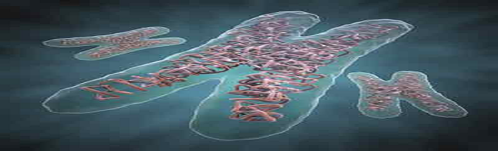
Enteroviruses are small viruses that are made of ribonucleic acid (RNA) and protein. This group includes the polioviruses, coxsackieviruses, echoviruses, and other enteroviruses. In addition to the three different polioviruses, there are over 60 types of non-polio enteroviruses that can cause disease in humans. Enteroviruses can be found in respiratory secretions (e.g., saliva, sputum, or nasal mucus) and stool of an infected person. Other persons may become infected by direct contact with secretions or stool from an infected person or by contact with contaminated surfaces or objects, such as a drinking glass or telephone. Parents, teachers, and child care center workers may also become infected by contamination of the hands with stool from an infected infant or toddler during diaper changes. Enteroviral disease is a common, under-recognized childhood illness. Most enterovirus infections during pregnancy cause mild or no illness in the mother. Although the available information is limited, currently there is no clear evidence that maternal enteroviral infection causes adverse outcomes of pregnancy such as abortion, stillbirth, or congenital defects. However, mothers infected shortly before delivery, may pass the virus to the newborn. Babies born to mothers who have symptoms of enteroviral illness around the time of delivery are more likely to be infected. Newborns infected with an enterovirus usually have mild illness, but rarely they may develop an overwhelming infection of many organs, including liver and heart, and die from the infection. The risk of this severe illness is higher for the newborns infected during the first two weeks of life. Each virus obtains its antigenicity from the capsid proteins surrounding the RNA core. According to the Centers for Disease Control and Prevention (CDC), 66 human serotypes of enteroviruses have been identified; however, a small number cause most outbreaks. Since the implementation of polio vaccines, the incidence of wild type polio has been eradicated in the Western hemisphere.
The most common form of human transmission is the fecal-to-oral route. Although respiratory and oral-to-oral routes are possible, they are more likely to occur in crowded living conditions. Enteroviruses are quite resilient. They remain viable at room temperature for several days and can survive the acidic pH of the human gastrointestinal tract. The incubation period is usually 3-10 days. Humans may be infected by direct contact of infectious material with broken skin or mucous membranes, accidental parenteral inoculation or aerosol. Diagnosticians collecting samples should also take the appropriate precautions.
Taqman Real time PCR assay.
• Use Real time PCR when enteroviral infection suspected, especially for neurologic infections.
• RT-PCR method is standard of care for diagnosing viral infection from CSF specimen.
• Rapid turnaround time aids in clinical management of patient.
• Assess viral Load measured by changes in the Enteroviruses RNA levels.
• Assess prognosis and early diagnosis for better patient cure.
• Confirm active Enteroviruses infection In patient.
• Elevation of the levels of CSF protein in about 50% of cases.
• Suspected patient with evidence of a neurologic infection (such as encephalitis, meningitis, or
Acute flaccid paralysis persons.
• Residing in areas where extended community outbreaks exist.
• International travelers to regions of the world where Enteroviruses is endemic.
• Vaccinated patient.
• During pregnancy.
Every day.
3-4 days.
Blood, serum, plasma, Collect in: Lavender (EDTA), pink (K2EDTA), or serum separator tube. Cerebrospinal fluid (CSF) Stability collection to initiation of testing On Cells: Ambient: 4 hours; after separation from cells: Refrigerated: 48 hours; Frozen at -20°C: 72 hours; Frozen at -70°C: 4 months. Do not thaw avoid repeated freezing and thawing.
NOTE: Collect CSF specimen in sterile screw capped bottles under all aseptic precautions for attempting isolation of virus minimum 0.5ml of CSF is required for acceptance of standard diagnosis.
CSF, Separate serum or plasma from cells within 24 hours.
Frozen-20°C. Refrigerate specimens at 2°C-4°C.
Heparinized specimens, Hemolysis sample, Quantity not sufficient for analysis, specimen grossly contaminated, specimen too old, frozen whole blood specimen, specimen leaky or tube broken.
This test can quantitate/detect Enteroviruses RNA Virus over the linear range 90-108 copies/mL. However this does not mean that lower copies or higher copies cannot be detected. The lower copies can be detected in some cases. This is a limitation of the currently available extraction systems. A negative result does not preclude the presence of Enteroviruses infection because results depend on adequate/proper patient sample storage and transportation as RNA is fragile and thermo labile, absence of inhibitors and sufficient RNA to be detected. Enteroviruses are a viral disease, and as such, antibiotics are of no value in the treatment of the infection. There is no hyperimmune E globulin available for pre- or post-exposure prophylaxis. Use Real time PCR when enteroviral infection suspected, especially for neurologic infections. RT-PCR method is standard of care for diagnosing viral infection from CSF specimen and confirm disease occurrence. Viral culture less rapid and less sensitive than RT-PCR.
The result of this test must always be correlated with clinical status and history of the patient and other relevant data and should not be used alone for the interpretation.